Case 26: A 55-Year-Old Man in the Emergency Department
A 55-year-old man with end-stage renal disease, is in the emergency department complaining of severe weakness. He had been suffering from an upper respiratory infection with fever and severe cough and had failed to attend the hemo-dialysis clinic:
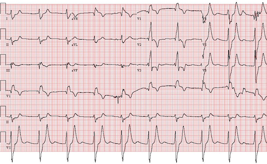
- Wide-QRS rhythm, 52/min. P waves are not recognizable
- Left axis deviation
- Right bundle branch block pattern. QRS duration 320 ms.
- Tall peaked T waves in the inferior and left chest leads.
- The ECG findings are consistent with severe hyperkalemia
Comments: The serum K was 9.2 mmol/L. (normal = 3.5 – 5.1 mmol/L) Hemodialysis was promptly started. Severe hyperkalemia slows atrial and ventricular conduction. When the K concentration rises above 7.0 mmol/L the P waves become smaller and eventually are no longer seen. The QRS becomes increasingly wide. The T wave also widens, however, its duration is usually not greater than that of the QRS (contrary to what happens in other forms of ventricular conduction abnormalities or ectopic ventricular arrhythmias in which the T wave is much wider than the QRS complex)

Hemodialysis was promptly started. The following ECG is recorded 9 minutes after ECG 1

- Wide QRS rhythm, 57/min. No P waves seen
- Right axis deviation
- Right Bundle Branch Block – Compared with the previous tracing, the QRS duration is less prolonged (208 ms)
- Tall peaked T waves in the left chest leads

Third ECG is recorded 14 minutes after ECG 1:

- Wide QRS rhythm – No P waves seen
- Right axis deviation
- Right bundle branch block – QRS duration 120 ms.
- Tall peaked T waves in V3 to V6

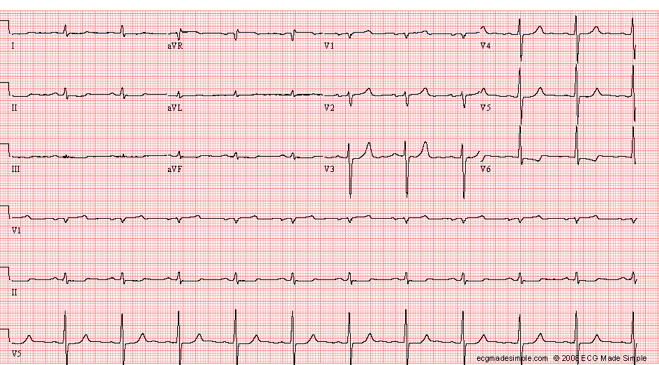
This ECG is recorded on the following day. Serum K was 4.1 mmol/L. The tracing was recorded at half standardization:

- Sinus rhythm, 66/min
- QRS duration 100 ms.
- Non-specific ST and T abnormality

ECG ID: E553