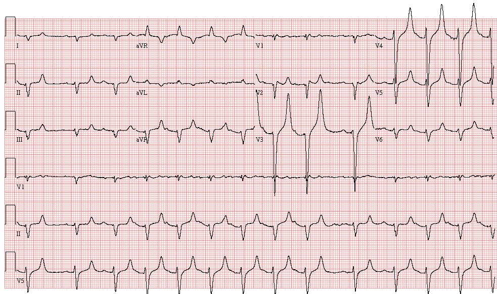
Case 35: An 83-Year-Old Man in Low-Output Failure
This 80-year-old man with known ischemic cardiomyopathy presents to the Emergency Department with severe low-output failure, hypotension, and hypoxemia. He denies any chest pain:
- Sinus rhythm, with a run of non-sustained atrial tachycardia, ventricular rate 80/min
- First degree AV block
- Nonspecific Intraventricular Block
- Septal infarction, age undetermined
- Very tall, symmetrical, peaked T waves in the chest leads, consider hyperkalemia
On bloodwork, her serum potassium was found to be 8.3 mmol/L (normal = 3.5-5.1 mmol/L)
A frequently asked question is: how do these tall T waves compare with those found in hyperacute ischemia/infarction? The answer to this is that the “hyperacute T waves” of ischemia/infarction are tall and symmetrical, but typically broad-based, not narrow and pointed (see ECG ID: E552 for an example of hyperacute T waves in the early stage of an acute anterior infarction).
In this severely ill patient with an ischemic cardiomyopathy, the possibility of “hyperacute T waves” was also considered in the differential diagnosis.
ECG ID: E658