Case 33: A 60-Year-Old Woman who Fainted
This 60-year-old woman is sent to the Emergency Dept. by her family physician, who had seen her after an episode of sudden-onset syncope, lasting several minutes. She reports a one month history of brief episodes of weakness and presyncope:
- Sinus bradycardia, 54/min
- Right bundle branch block (RBBB)
- Left anterior fascicular block (LAFB)
- Bifascicular block (RBBB and LAFB)

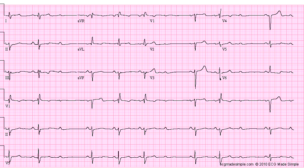
The patient is admitted to the cardiac ward and monitored. In hospital, the patient is symptom free. The following ECG is recorded the next day: 
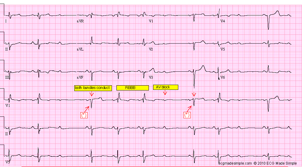
- Sinus rhythm, with Mobitz II second degree AV block – ventricular rate 46/min
- Right bundle branch block
- Left anterior fascicular block
- Bifascicular block
Comment: The QRS complexes following the longer R-R intervals caused by the AV block show only LAFB. Mobitz II second degree AV block is nearly always at the level of the His-Purkinje system, and is caused by bilateral bundle branch block. This case is a good illustration of the site of block. After the long RR interval, both bundles are able to conduct. As the right bundle has a longer recovery period than the left bundle, the next QRS complex shows a RBBB and LAFB pattern (the AV conduction is through the posterior fascicle of the left bundle which has recovered). Finally, both bundles fail to recover and AV block occurs. The PR interval does not change. The AV node appears to conduct normally.
After a therapeutic procedure, the following ECG is obtained:
- DDD pacing (atrial sensed, ventricular paced rhythm)
- Sinus rhythm, 60/min

ECG ID: E663