Case 105: A 72-Year-Old Man complaining of Weakness
A 72-year-old hypertensive diabetic man is brought to the Emergency Department because of nausea, malaise and increasing weakness for the past 3 days. He had very little fluid intake, despite very hot humid weather. He had continued to take his medication, that included spironolactone and indomethacin:
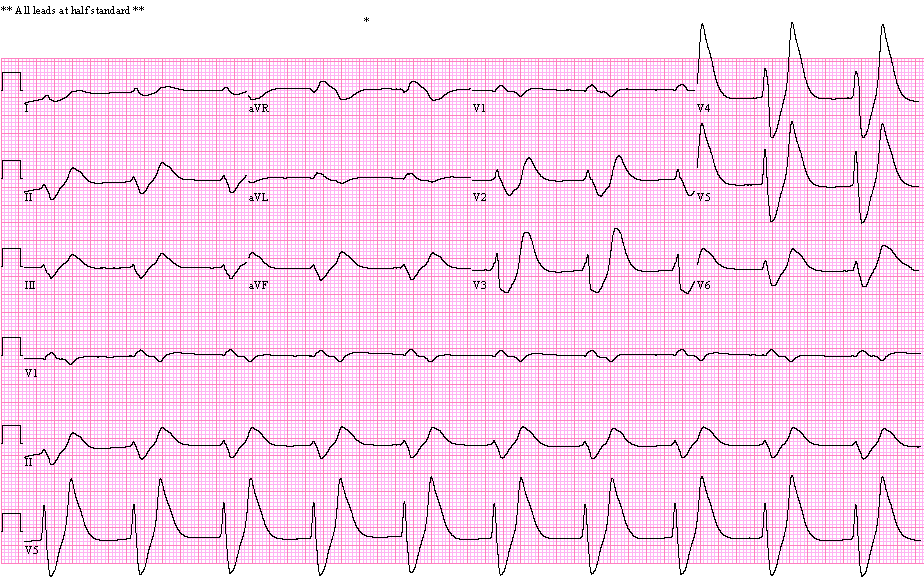
- No P waves are seen
- Wide QRS rhythm, 57/min (QRS duration = 235 msec)
- RBBB pattern / Tall T waves
- Consider hyperkalemia
The rhythm is regular, 57/min. No P waves are seen. The QRS is very wide and bizarre. The width of the tall T waves is very close to that of the QRS. (In intraventricular conduction abnormality due to other etiologies and in ventricular arrhythmias, such as idioventricular rhythms, the T wave is significantly wider than the QRS). All these findings are strongly suggestive of hyperkalemia.
The serum Potassium of this patient was 10 mmol/L (normal = 3.5 – 5.1 mmol/L)
Reference: Parham WA, Mehdirad AA, Biermann KM, Fredman CS. Hyperkalemia revisited. Tex Heart Inst J. 2006;33(1):40-7. PMID: 16572868; PMCID: PMC1413606. https://www.ncbi.nlm.nih.gov/pmc/articles/PMC1413606/

The patient received IV calcium chloride, insulin, sodium bicarbonate. He underwent dialysis. This ECG was recorded 1 1/2 hours after ECG 1. The serum K was 6.4 mm/L:
- Atrial fibrillation, 70/min
- Moderate voltage criteria for left ventricular hypertrophy ( S in V1 + R in V6 = 35 mm, R in V5 30 mm)
- Tall peaked T waves
Evolutionary changes often seen in severe hyperkalemia during treatment.
ECG is recorded one day later. The serum K was 4.6 mmol/L:
- Sinus rhythm, 75/min
- Normal ECG
The patient made a good recovery and was discharged from hospital.
ECG ID: E248